Abnormal glycosylation is a hallmark of cancer. Several carbohydrate structures are highly expressed in cancerous cells compared to normal tissue, including the Tn (GalNAcα1-O-Ser/Thr), STn (Neu5Acα2-6GalNAcα1-O-Ser/Thr) and T (Galβ1,3-GalNAcα1-O-Ser/Thr antigens (1,2). Antibodies to these can act as markers for cancerous cells (3) and have been used as the platform for diagnostics, therapeutics, and anti-cancer vaccines (4-6).
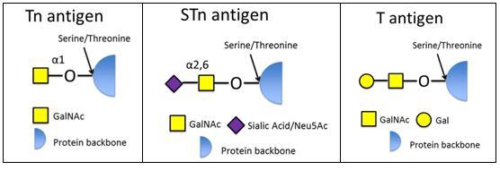
The Tn, STn and T antigen are often components of mucin protein, which are upregulated in cancerous cells (7). Mucins are large glycoproteins, either secreted from or membrane-bound in epithelial cells, in which O-linked oligosaccharides comprise more than 50% of the molecule by weight (8). They are up-regulated in cancerous cells and their localization changes from being on the apical side of normal epithelium to symmetrically distributed in malignantly transformed epithelium (Bos). Mucins are often abnormally glycosylated in cancerous cells and tissues (3, 7-9).
- Hakomori, S. (1996) Cancer Res. 56, 5309
- Springer, G.F. (1984) Science 224, 1198
- Lavrsen, K. (2012) Glycoconj. J., DOI 10.1007/s10719-012-9437-7
- Desai, P. (2000) Transfusion Med. Rev. 14, 312-25
- Ingale, S. et al. (2007) Nat. Chem. Biol. 10, 663
- Miles, D. et al. (2006) J. Biol. Chem. 281, 3586
- Taylor-Papadimitriou, J. (1999) BBA 1455, 301
- Brockhausen, I. (2006) EMBO Reports 7, 599
- Van Elssen, C.H.M.J. (2010) Histopathology 57, 597
SBH Sciences is looking for partners to investigate our glycosyltransferase enzymes and carbohydrate antibodies as diagnostic tools, therapeutics or for production of glycol-peptide/glycol-protein products. Moreover, our extensive bioassay and in vitro biomarker analysis capabilities are available to support development of your product.
We are open for suggestions and would be pleased to hear from you.
Our expertise is your competitive edge!
Anti-Tn Antibody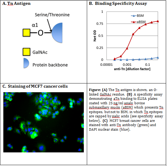
Description:
Dysregulation of cellular glycosylation is a common characteristic of many cancers (1). The Tn antigen (GalNAcα1-O-Ser/Thr), is up-regulated in a wide variety of cancers (2,3), and has been investigated as a diagnostic & prognostic marker, and as a therapeutic (4). It has also been explored as a component of anti-cancer vaccines (5). The specificity of aTn was shown by binding to asialo bovine submaxillary mucin (aBSM), which presents the Tn antigen, and not to BSM in which the Tn epitope is shielded by sialic acid residues (ref 3 and Figure 1 panel B). This antibody has previously been sold under the name HB-Tn (6,7).
- Hakomori, S. (1996) Cancer Res. 56, 5309.
- Springer, G.F. (1984) Science 224, 1198.
- Kjeldsen, T. et al. (1988) Cancer Res. 48, 2214.
- Desai, P. (2000) Transfusion Med. Rev. 14, 312-25.
- Ingale, S. et al. (2007) Nat. Chem. Biol. 10, 663.
- Marcos, N.T. et al. (2004) Cancer Res. 64, 7050.
- Julian, S. et al. (2001) Glycocon. J. 18, 883.
Preparation: Monoclonal antibodies were produced from mouse hybridoma 5F4 clone.
Purification: Three step liquid chromatography, proprietary.
Catalog #: SBH-Tn
Formulation: Sterile filtered solution of PBS at 0.5mg/ml.
Stability: Stable for 4 weeks at 4C. Stable for 6 months at -80C. Avoid repeated freeze-thaw cycles.
Purity: Greater than 90% by SDS-PAGE.
Reconstitution: N/A
Specificity Assay: To create asialo bovine submaxillary mucin (aBSM), 30mg of BSM (Sigma M3895) was treated with 5U of Neuraminidase (Sigma N2876) for 2 hrs at 37⁰C. Double dilution ELISAs were carried out by coating 96 well plates with a 1:2 serial dilution of 10ng/ml BSM or aBSM, blocking and incubating with a 1:4 serial dilution of anti-Tn (SBH-Tn). After washing, plates were incubated with a goat-anti-mouse (IgG+IgM)-HRP conjugate (Thermo 31446) at 1:1000 dilution. After washing, plates were developed with 2x ABTS (Southern Biotech 0202-01). Net OD was measured at 405 – 650 nm. SBH-Tn MAb diluted 4,000 fold could detect aBSM coated at 25 ng/ml, with 30 fold less signal for BSM (see Figure 1 panel (B) above).
Usage: For research only. Not for use in diagnostic or therapeutic procedures.
Country of Origin: USA
To learn more about the Tn, STn, T and other carbohydrate-based, cancer-specific antigens, please visit the Carbohydrate Cancer Antigens page.
Anti-STn Antibody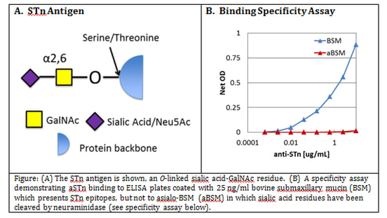
Description:
Dysregulation of cellular glycosylation is a common characteristic of many cancers (1). The STn antigen (Neu5Acα2-6GalNAcα1-O-Ser/Thr), also known as the sialyl-Tn antigen, is up-regulated in a wide variety of cancers (2). It is formed from the Tn antigen (GalNAcα1-O-Ser/Thr) through the action of the ST6GalNAc family of enzymes (3,4). STn-Keyhole Limpet Hemocyanin conjugate has been tested as an anti-cancer vaccine (5). The specificity of the anti-STn monoclonal antibody was shown by binding to bovine submaxillary mucin (BSM), which presents the STn antigen, and not to asialo-BSM (ref 2 and Figure 1 panel B). This antibody was previously marketed under HB-STn (3,4).
- Hakomori, S. (1996) Cancer Res. 56, 5309.
- Kjeldsen, T. et al. (1988) Cancer Res. 48, 2214.
- Marcos, N.T. et al. (2004) Cancer Res. 64, 7050.
- Julian, S. et al. (2001) Glycocon. J. 18, 883.
- Miles, D. et al. (2006) J. Biol. Chem. 281, 3586.
Preparation: Monoclonal antibodies were produced from the mouse hybridoma 3F1 clone (HB-STn).
Purification: This IgG1 was purified by Protein G affinity chromatography.
Catalog #: SBH-ASTn
Formulation: Sterile filtered solution PBS, at a stock concentration of 640 ug/mL.
Stability: Stable for 4 weeks at 4C. Stable for 6 months at -80C. Avoid repeated freeze-thaw cycles.
Reconstitution: N/A
Specificity Assay: To create asialo bovine submaxillary mucin (aBSM), 30mg of BSM (Sigma M3895) was treated treated with 5U of Neuraminidase (Sigma N2876) for 2 hrs at 37⁰C. Double dilution ELISAs were carried out by coating 96 well plates with a 1:2 serial dilution of 10ng/ml BSM or aBSM, blocking and incubating with a 1:4 serial dilution of 10ug/ml anti-STn (SBH-STn). After washing, plates were incubated with a goat-anti-mouse (IgG+IgM)-HRP conjugate (Thermo 31446) at 1:1000 dilution. After washing, plates were developed with 2x ABTS (Southern Biotech 0202-01). Net OD was measured at 405 – 650 nm. As low as 2ng/ml SBH-STn MAb could detect BSM coated at 25 ng/ml, with no detectable binding to aBSM (see Figure 1 panel (B) above).
Usage: For research only. Not for use in diagnostic or therapeutic procedures.
Country of Origin: USA
To learn more about the Tn, STn, T and other carbohydrate-based, cancer-specific antigens, please visit the Carbohydrate Cancer Antigens page.
Anti-T Antibody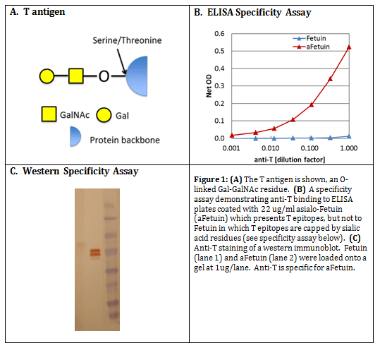
Description:
Dysregulation of cellular glycosylation is a common characteristic of many cancers (1). The T antigen (Galβ1,3-GalNAcα1-O-Ser/Thr ) is up-regulated in a wide variety of cancers (2, 3), and has been investigated as a diagnostic & prognostic marker, and as a therapeutic and vaccine (4, 5). The specificity of the anti-T monoclonal antibody was shown by binding to asialo-fetuin, which presents the T antigen, and not to fetuin, in which the T antigen is capped by sialic acid residues (3). This antibody was previously marketed under HB-T (6).
- Hakomori, S. (1996) Cancer Res. 56, 5309.
- Springer, G.F. (1984) Science 224, 1198.
- Kjeldsen, T. et al. (1988) Cancer Res. 48, 2214.
- Desai, P. (2000) Transfusion Med. Rev. 14, 312-25.
- Springer, G.F. (1997) J. Mol. Med. 75, 594.
- Marcos, N.T. et al. (2004) Cancer Res. 64, 7050.
Preparation: Monoclonal antibodies were produced from the mouse hybridoma 3C9 clone (HB-T).
Purification: Three step liquid chromatography, proprietary.
Catalog #: SBH-T
Formulation: Sterile filtered solution PBS.
Stability: Stable for 4 weeks at 4C. Stable for 6 months at -80C. Avoid repeated freeze-thaw cycles.
Reconstitution: N/A
Specificity Assay: To create asialo Fetuin (aFet), 5mg of Fetuin (Sigma F3004) was treated with 2U of Neuraminidase (Sigma N2876) for 2 hrs at 37⁰C. Double dilution ELISAs were carried out by coating 96 well plates with a 1:3 serial dilution of 67ug/ml Fet or aFet, blocking and incubating with a 1:3 serial dilution of anti-T (SBH-T). After washing, plates were incubated with a goat-anti-mouse (IgG+IgM)-HRP conjugate (Thermo 31446) at 1:1000 dilution. Plates were developed with 2x ABTS (Southern Biotech 0202-01). Net OD was measured at 405 – 650 nm. SBH-T MAb diluted 10 fold could detect aFet coated at 22 ug/ml, with 200 fold less signal for Fet (see Figure 1 panel (B) above).
Usage: For research only. Not for use in diagnostic or therapeutic procedures.
Country of Origin: USA
To learn more about the Tn, STn, T and other carbohydrate-based, cancer-specific antigens, please visit the Carbohydrate Cancer Antigens page.
Glycosyltransferase Enzymes
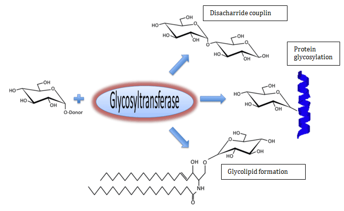
Glycosyltransferase enzymes add monosaccharides onto sugars, proteins, and lipids with perfect specificity. Over half of the proteins expressed in the human genome undergo post-translational glycosylation, which is accomplished through the coordinated activity of hundreds of glycosyltransferases. Glycosylation affects a wide variety of protein functions, including activity, resistance from protease cleavage, and half-life in circulation.
Glycosyltransferases catalyze the transfer of monosaccharides from activated donors, frequently nucleotide sugars, to acceptor substrates. Shown in the figure, glucose is transferred from UDP-glucose to an acceptor.
The half-life of protein-therapeutics in circulation can be dramatically altered by changing the glycosylation state, due to increased clearance by the kidney or hepatic receptors (Varki et al. (2003) "Essentials of Glycobiology", 2nd edition, Cold Spring Harbor Lab. Press). For example, removal of all sialic acid residues from erythropoietin (EPO) to form asialoerythropoietin decreases it's circulation half-life by more than 150 fold (Erbayraktar et al. (2003) Proc. Nat. Acad. Sci. 100, 6741).
Glycosylation can also confer resistance to protease-cleavage-inactivation of proteins. Fibroblast growth factor 23 (FGF-23) is implicated in familial tumoral calcinosis (FTC), a metabolic disorder. One of the causes of FTC is lack of O-glycosylation of FGF-23, which renders it susceptible to cleavage and unable to be secreted (Kato et al. (2006) J. Biol. Chem. 281, 18370). Low-density lipoprotein receptor is also protected from cleavage by O-glycosylation in the extracellular stalk region, and lack of glycosylation results in the loss of the receptor region (Kingsley et al. (1986) J. Biol. Chem. 102, 1576).
Glycosylation of lipids is also important for a wide variety of purposes. The glycolsphingolipid gangliosides act as intracellular signaling molecules, and glycosylphosphatidylinositol can act as membrane attachments for proteins (GPI anchor).