
SBH Sciences is offering In-Vitro models for Inflammation and Fibrosis [NASH]
SBH Sciences is offering several In-Vitro inflammation models. The first model is based on utilization of human PBMC and the secretion of human TNF-alpha as a result of LPS stimulation.
Innovative Models
Liver (LX-2) / Lung (Beas-2B) / Heart models (induced pluripotent stem cells - iPSC)
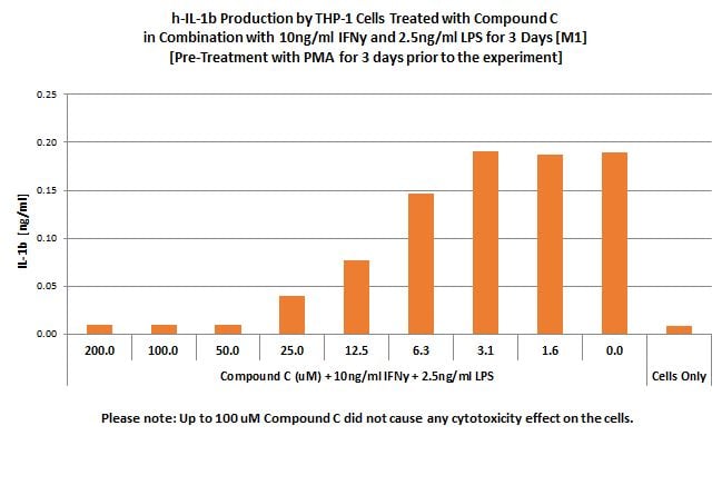
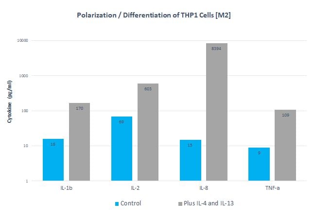
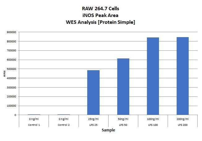
- Macrophages activation M1 & M2
- Functional assays (receptors-ligands interaction)
- Inflammatory Cytokines and Fibrosis Biomarkers
- Cytotoxicity – apoptosis
Biomarkers Platforms
- ELISA / Receptor Binding Assay for biomarkers and metabolites
- Luminex / MSD / ELLA – multi-array biomarkers
- PeggySue, WES and Jess
- Flow cytometer [FACS]
- Isolight
- qRT-PCR
Markers (partial list)
TNF-a, MCP-1, IL-1b, TGF-beta, iNOS, Pro-Collagen, Galectin-3, TLRs, Integrins
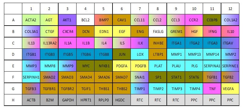
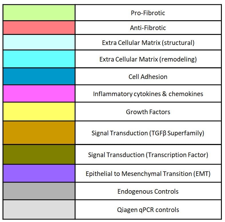
mRNA Analysis of Pro-Fibrotic Chemokines / Cytokines / Collagen for up and down-regulation in Fibrosis Model
Effect of GR-MD-02 on cell culture model of macrophages: THP-1 cells treated with phorbol ester and endotoxin
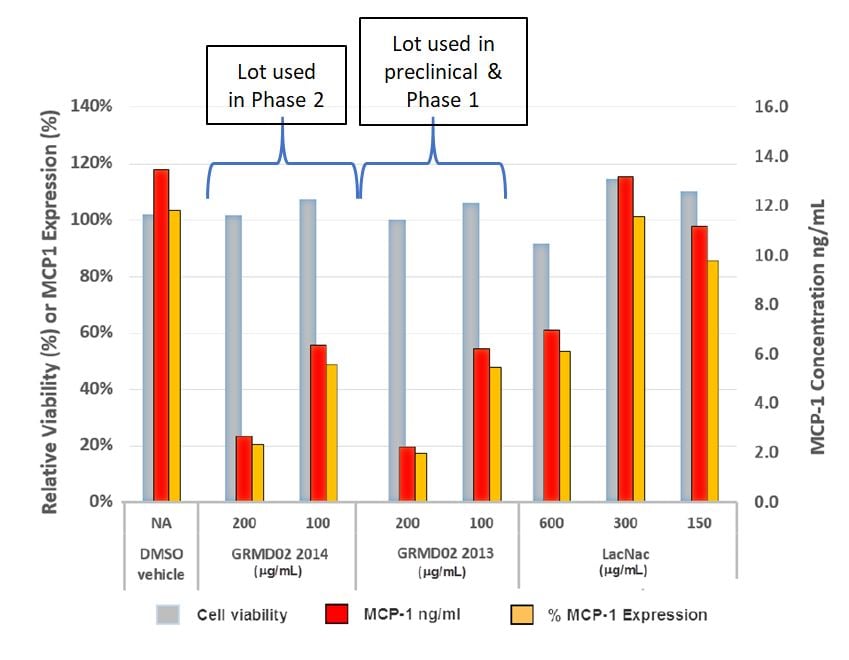
Expression of MCP-1 is reduced in dose dependent fashion in two separate GMP lots of GR-MD-02 while viability of THP-1 cells is maintained (compared to N-lactosamine—LacNac)
Note: We have shown that Galectin-3 is expressed in THP-1 cells. There is also a reduction in TNF-alpha and IL-8 with treatment.
Contact SBH
Please contact us with your specific needs or any questions related to In-Vitro Inflammation investigation and anti-inflammation lead drug optimization. We can also analyze all your animal studies samples using our innovative bioanalytical platforms.